


יום פתוח בטכניון
בואו להכיר מקרוב את הטכניון ביום הפתוח ב-5/3 בקמפוס הטכניון בחיפה


שיתוף פעולה ייחודי נרקם בטכניון: צעירים מבית הלוחם בחיפה הצטרפו לקורס מעשי בפקולטה להנדסת ביוטכנולוגיה ומזון, ובסיומו הציגו המשתתפים בירות שייצרו בעצמם במרכז קרסו לחדשנות בטכנולוגיות מזון, שנחנך לאחרונה בטכניון

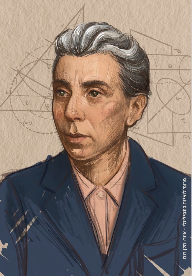
פרופ' דבי לינדל מהפקולטה לביולוגיה זכתה בפרס אוולין האצ'ינסון באוקיינוגרפיה והתקבלה לאקדמיה האמריקאית למיקרוביולוגיה

הרצאתו של ד"ר יותם בר-און מהפקולטה לרפואה ע"ש רות וברוך רפפורט : מדוע הנגיפים תמיד מנצחים?

הכנס הישראלי השני לפילוסופיה של בינה מלאכותית
04.03.2026 רביעי, בשעה 09:30
הוספה ליומן

יום פתוח בטכניון 5.3
05.03.2026 חמישי, בשעה 09:30
הוספה ליומן
תערוכת "מראות מקום"
30.11.2025 ראשון, בשעה 09:00
הוספה ליומן
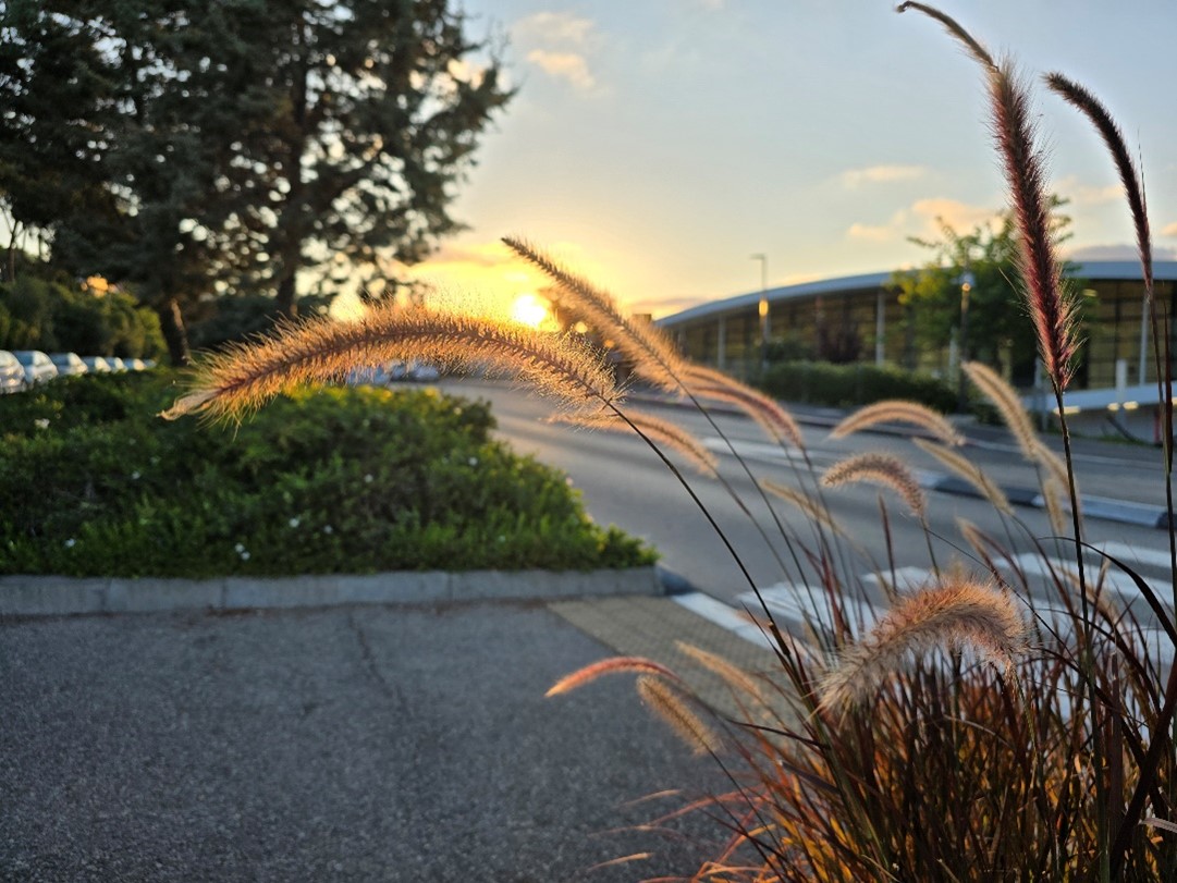
תערוכת הצילום "טבע בקמפוס"
16.07.2025 רביעי, בשעה 09:00
הוספה ליומן
100000
בוגרים
18
פקולטות
15000
סטודנטים
60
מרכזי מחקר
ברחבי הקמפוס